Description
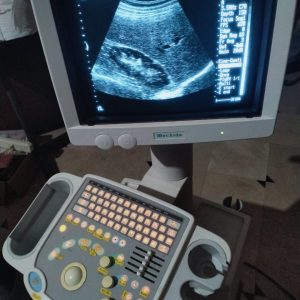
The SONOLINE G20™
- All-digital signal processing technology creates excellent image quality
- State-of-the-art beam forming provides a wide acoustic aperture, improved image resolution, uniformity and penetration for difficult-to-image patients
- All-digital G20 system architecture enables seamless integration of advanced capabilities including DICOM connectivity solutions, Tissue Harmonic Imaging (THI) and TGO™ tissue grayscale optimization technology
- Ongoing performance enhancements, which require only simple, cost-effective software upgrades, protect your initial investment into the future
The SONOLINE G20 ultrasound system goes beyond excellent image quality to meet your clinical needs. The G20 system incorporates superb workflow advancements, user matched ergonomics and proven Siemens reliability. The results are efficiency, time savings and greater patient throughput. Without interruption.
- Slim, lightweight design with small footprint and four swivel wheels makes the system ideally suited for use in tight work spaces and easily maneuverable from suite to suite and patient to patient
- Intuitive interface with the familiar Windows®-based operating system promotes efficient and ergonomic system control via on-screen menus and proprietary ReadySet™ workflow shortcuts
- QuickSet™ user programmable system parameters and integrated cine review streamline exams and enhance workflow
- DIMAQ-IP integrated workstation with built-in CD-R/W drive allows quick and easy digital acquisition, storage, review, quantification and transfer of complete ultrasound studies
- TGO technology for instantaneous, one-button image optimization
- Ergonomically designed transducers incorporate lightweight microCase™ transducer miniaturization technology to promote user comfort
- Embedded connectivity solutions allow simple integration into DICOM-enabled networks and PC-based workstations
- Next-generation all-digital technology for unparalleled grayscale imaging performance in a compact, ultra-portable system
- Superb system ergonomics deliver excellent workflow efficiencies to save time in busy clinical settings
- DIMAQ-IP integrated workstation with built-in CD-R/W drive promotes efficient and cost-effective output and archiving of exam data
- Tissue Harmonic Imaging (THI) is available to enhance visualization, particularly in difficult-to-image patients
- MultiHertz™ multiple frequency imaging optimizes penetration and resolution to expand the clinical versatility of each transducer
- Software upgradeability expands and protects your initial investment today and into the future
Specifications
Physical System Specifications
- Height 128.3 cm (50-5 inch)
- Width: 46.4 cm ( 18.3 inch)
- Depth: 67.5 cm (26.6 inch)
- Weight: 60 kg (132 pounds) without OEM’s
Monitor
- Monochrome, 12-inch, high resolution, progressive scan (non-interlaced) CRT
- 640 x 480 pixel display matrix with a recordable image area of 640 x 480 pixels (VGA)
- Monitor tilt of 17 degrees up, 17 degrees down and swivel of 150 degrees right and 150 degrees left
- Brightness and contrast controls
User Interface
- User-centric control panel with home base layout
- Intuitive personal computer operating principles
- ReadySet on-screen workflow shortcuts for instant access to most frequently used functions
- On/Off task light and back-lit illumination of control panel
- On Screen Menu (OSM) provides easy and immediate access to secondary imaging controls
- Wrist support to help reduce operator repetitive stress disorders
- Alphanumeric keyboard for standard text, function keys and system programming
- Up to 32 user-definable QuickSet user- programmable system parameters for individual transducer/application settings. QuickSets combine all preferred imaging mode parameters, annotation text and measurements into one user preset
- Configurable two-pedal footswitch
Operating/ Display Modes
- 2D imaging in fundamental and harmonic modes
- Flexible combination of imaging modes in side-by-side Dual and Dual Select in real-time and digital cine replay
- Selectable split screen display formats in 2D with M-mode: top-bottom or side-by-side in real-time and digital cine replay
- Tissue M-mode
Hard Drive
- Internal 40 GB Hard Drive
- Image storage capacity up to 42,000 black/white images
- Storage of approximately 6,300 dynamic clips (option)
CD-R/W Drive
- Supports 650 MB or 700 MB read/write CD-R
- Allows storage and archiving of complete patient studies including still images, dynamic clips (optional), reports and measurements Storage capacity dependent upon patient study size
- Export of images and clips (optional) in TIFF and AVI file format, DICOM exchange Media (optional)
Power Supply
- 115V/230V version: 98-132 VAC, 50/60 Hz/196-264 VAC, 50/60 Hz
Image File Format
- Standard output: TIFF & DICOM
Patient Study Management
Replay of digitally stored images in a select-able 1-up, 4-up, 9-up or 16-up screen format. The patient study screen allows for study selection, search, and deletion or for export to CD-R.
- CD-R/W drive for 650 and 700 MB CD-R provides patient study archiving
- Storage and retrieval of frozen static images
- Storage and retrieval of reports
- Instant dial-in replay of static images in 1-up screen format
- Perform Measurements and Calculations on current, as well as on saved and retrieved images
- Export of patient studies from Hard Drive to CD-R/W Drive
- Exported images in a standard TIFF format
- Patient database sorting by Name, ID, Study Date and Exam type
System Applications & Reporting
- Abdomen
- Emergency Medicine
Pain Management Imaging
- Orthopedic
Women’s Imaging
- Breast Imaging
- Gynecology
- Obstetrics
Small Parts
- Breast
- Testicle
- Thyroid
Urology Imaging
- Pelvis
- Prostate
Vascular Imaging
- Transcranial
Transducer Ports
- Up to three transducers can be connected simultaneously
- Two universal transducer ports that support curved array and linear array
- One mechanical sector array port (optional)
- Electronic transducer selection (instantaneous switching between transducers)
- Parking port for convenient storage of a third transducer connector (LC system configuration)
- Industrial design provides easy access to the transducer ports
Transducers/ Probes
- Linear Array 5-10 MHz*
- Curved Array 2-8 MHz*
- Endocavity 4-9 MHz
- Endovaginal 4-9 MHz
- Bi-plane Endorectal 5-7.5 MHz
Transducers:
- L10-5
- 7.5L75s
- C8-5
- C5-2
- C4-2
- EV9-4
- EC9-4
- ENDO PII


Reviews
There are no reviews yet.