Description
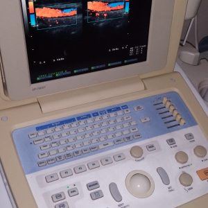
Full Digital Ultrasound System
UF-870AG
An Intelligible & Premium Ultrasound System
By featuring Fukuda Denshi’s original F-AG technologies in its compact body,
the UF-870AG ensures excellent image quality and high operational efficiency.
With a variety of advanced functions, this revolutionary full digital
ultrasound system initiates a new high-quality imaging environment.
Advanced Image
Advanced Function
Advanced Interface
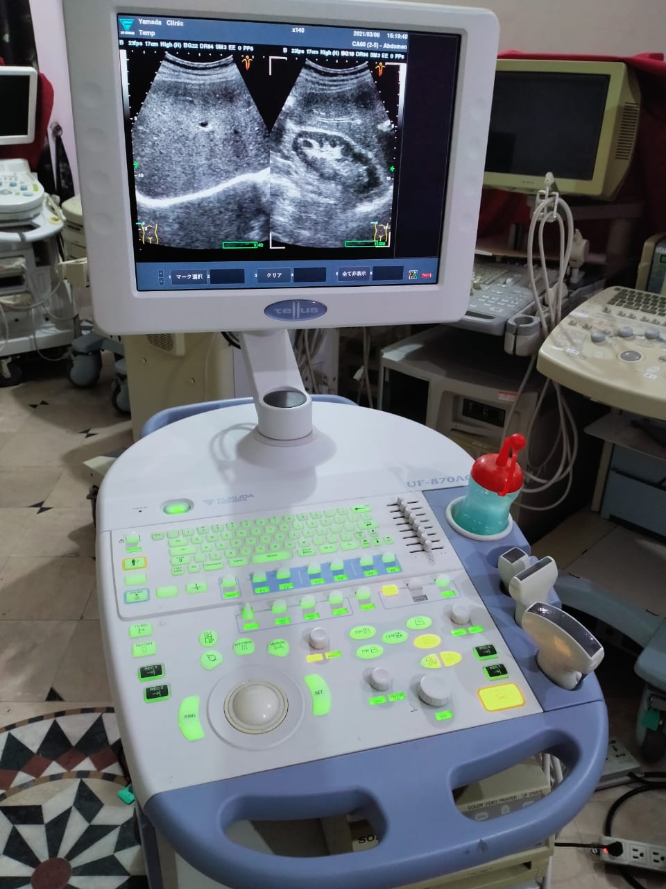
The built-in digital beam former provides 4 x 64 channels, enabling
simultaneous reception of the images in four directions. It also realizes an
ever high frame rate and improves the scanning line density.
Increasing the number of receiving signals improves
the color Doppler frame rate.
This new function enables the adjustment of the frequency
to an optimum for field depth and applications.
This ensures reduced examination time and improved workflow.
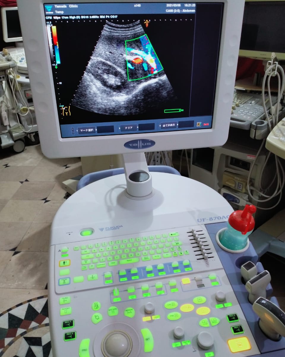
The digital beam processing enables the detection of delicate blood
flow (none missed).
And by mixing every received signal, a Doppler color flow mapping
of sensitivity 1.5 times higher than a conventional system is produced,
thereby increasing diagnostic efficacy.
Optional functions are available in abundance, enabling
configuration of a system that exactly satisfies the need
of each individual user. Selection of optional functions
enables the user to use the UF-870AG as a system dedicated
to cardiovascular or abdominal/vascular application.
And also, it incorporates all the optional functions, which
can make the UF-870AG a multifunctional ultrasound system.
The UF-870AG incorporates a TFT LCD color monitor developed
for the ultrasound system that requires ultra-wide angle of
visibility and high brightness. This ensures to display highly
precise images in either bright or dark environment. The LCD
monitor position can freely be adjusted in any directions.
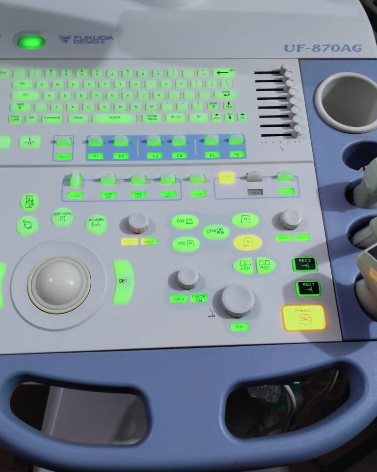
The operation panel can be slid left-right. Thus, an optimum
examination environment is made available in a limited space.
Keys are ergonomically laid out and properly labeled to
enable quick and efficient examination.
Network (option)
DMAS(Data Management and Archiving System)
Advanced Diagnosis
PC
For smooth examination flow
Network system enabling
total data management
Patient data management
Patient information, measurement results and ultrasound images are saved in the hard disk,
enabling easy data search and comparison with past data.
MWM
Movie data and still images
Still images
DICOM server
Printer
Reporting
A template is provided to enable the user to easily
compile the report in A4 size. Any PDF image can
simply be dragged into the report while comment(s)
can easily be entered. Also, a laser printer can be
directory connected to the UF-870AG to print out the
report
Display Unit 15-inch, High-Resolution, Ultra-wide view, TFT LCD monitor
Scanning methods Electronic Phased array / Convex array / Linear array
Beam Former Digital, Multi-Beam Processing
Frame Rate Max 224 frame/sec (depend on system setting)
Operation Mode B (2D) mode, M mode, PWD mode, CFM mode, Power Doppler mode, Triplex mode,
Color M mode, CWD mode (option), Anatomical M mode (option), Doppler Tissue Imaging (option)
B-mode Focus method Transmitting: 1 step (8 types selectable)
Receiving: continuous dynamic focus
Display Depth 2 ~ 30cm (depending on probe)
Frequency 2 types selectable and AFA (Automatic Frequency Adjustment)
Tissue Harmonic Imaging on/off, 2 types frequency selectable (depending on probe)
Color Scale Imaging 8 types B color selection
Display Control up/down, right/left, view angle variable (depending on probe)
M-mode Display mode Moving Bar
Sweep Speed 4 steps (2 to 8 sec/screen)
Color Scale Imaging 8 types M color selection
PWD mode Display Mode PWD mode, High PRF mode, Spectral Doppler Tissue Imaging (option)
Sweep Speed 4 steps (2 to 8 sec/screen)
Velocity range ± 1KHz to ± 20KHz
CFM mode Display Mode Velocity mode, Power mode, Doppler Tissue Imaging (option)
Velocity range ± 250Hz to ± 20KHz
Color Map Velocity: 4types, Power: 4types, DTI: 4types
CWD mode (option) Display method CWD mode, Pencil CWD mode
Sweep Speed 4 steps (2 to 8 sec/screen)
Velocity range ± 1KHz to ± 48KHz
Imaging Control GAIN 60 ~ 100dB, 32 steps
STC 8 steps, slide volume
Dynamic Range 48 ~ 96dB, 8 steps
Echo Enhance 8 steps
Frame Correlation 4 steps
Post Process 8 steps
Line Density 2 steps
Color Scale Imaging 8 types
ECG synchronized freeze Dual Time
Probe Connection 3 active connectors, 1 Pencil probe connector
Biological Signal (option) ECG, PCG
Measurements and
General, Cardiology, Vascular, Urology, OB/GYN measurement, with Report Function Calculations
Image Filing 600 frames Cine memory (max 3000 frames: Long Cine option)
USB memory, 160GB HDD, CD-R/RW
Network LAN, DICOM (option)
General Power AC100 ~ 240V ± 10%, 50/60Hz
Power Consumption Approx. 850VA
External Dimensions (W) 480 × (D) 826 × (H) 1315~1505mm
Weight Approx. 100kg
- Option Probe Lineup
- Biopsy Needle Guide Holders
- FUT-SA162-5A FUT-1-5PA FUT-3-8PA
- FUT-CA602-5A FUT-4-9MC FUT-TVA114-7A
- FUT-LA385-12A
- FUT-PAD60C for FUT-CA602-5A
- FUT-PAD50B for FUT-5-12L50
- FUT-PAD16A for FUT-SA162-5A
- FUT-PVG11A for FUT-TVA114-7A
- 652-014 for FUT-4-9MC
- 612-085 for FUT-LA385-12A
- FUT-5-12L50
- FUT-3-8TEM
- FUT-PEN2 FUT-PEN4 FUT-PEN8o Convex
- Linear
- Software Options
- Name Description
- UF-870AG-CWD CW Doppler
- UF-870AG-CPG Cardiac package (Tissue Doppler & Anatomic M-mode)
- UF-870AG-SCU DICOM store SCU
- UF-870AG-MWM DICOM modality worklist
- UF-870AG-STE Stress echo package
- UF-870AG-LCL Long cine option
- Other Options
- Name Description
- OA-293 Footswitch
- UF-800XTD-CMG Cable manager
- UF-870AG-RTY Recorder tray
- UF-870AG-ECG ECG unit
- MA-300 PCG microphone

Reviews
There are no reviews yet.